
Glioma Case study 1
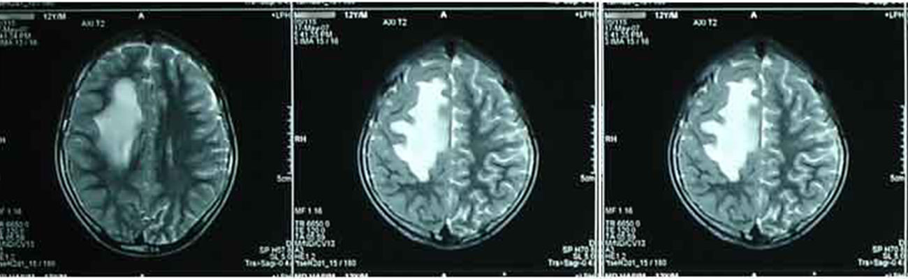
Md H. 12 years aged young boy came to our clinic on 16th of December 2008 with the complaints of seizures and headache off and on with haziness of vision, started since last 2 months.
MRI of BRAIN done on dated 4th of December 2008:- Nodular lesion with perifocal oedema in right sup parietal region ? Tuberculoma. Astrocytoma.
Stereotactic biopsy done on 10th of December 2008 showed "…High grade neoplasm ... Glioma..."
After undergoing treatment from us with the medicines Ruta 6c, two doses a day, Calcarea Phosphorica 3X, two doses a day, and Lycopodium 30c two doses daily for edema, all his clinical symptoms recovered within 6-7 months. Now the patient is leading a trouble free, normal life but he still continuing his medication in reduced doses.
Follow up M.R.I. of Brain done on dated 10th of January 2013: --"within normal limit."
Glioma Case study 2
Mrs. M.C. female, 50 years, came to us on 19th July, 1994 and presented with Giddiness; diminished hearing in the right side; slurring of speech; fear of falling down;
unbalanced gait; progressive weakness of the left side and forgetfulness. Her trouble of trembling of whole body had started the last 4 months.
C.T. Scan of Brain (plain and contrast study) done on 14th July, 1994 showed "…a fairly large infiltrating type ill-defined enhancing soft tissue mass lesion from the
apex of the right petrous bone with erosion of the apex of the petrous and adjacent base of skull and with intracranial extension in posterior
fossa and right temporal area as well as extension in right temporal fossa associated with moderate degree of obstructive hydrocephalus Gliomas"
After undergoing treatment from us with the medicines Ruta 6c two doses a day, Calcarea Phosphorica 3X two doses a day, all her clinical symptoms
had improved within 6 months. Now the patient is living her normal, trouble free daily life without any medication.
C.T. Scan of Brain done on 22nd November, 1994 “….As compared to the previous CT Scan done on 14th July, 1994 the almost identical appearance noted in the soft tissue mass at the right CP angle and adjacent base of skull area with bone erosion.
Follow up C.T. Scan of Brain done on 1st December, 1995, "…as compared to last CT Scan done on 22nd November, 1994 the infiltrating soft tissue mass adjacent to right petrous bone shows almost identical appearance…"
We then reduced the doses and stopped our treatment after one year.
After 9 years without any medicines follow up C.T.Scan of Brain (plain and contrast study) done on 6th August, 2005 shows "…A locally invasive soft tissue mass seen in relation to right petrous temporal bone which shows no significant change in comparison to last scan at 1st December, 1995."
After 12 years without any medication again Follow up C.T. Scan of Brain (plain and contrast study) done on 18th April 2008 Impression – CT findings in a follow-up case of right CT angle and temporal region irregular enhancing mass now showing almost identical position and size as compared to previous CT Scan done on 6th August 2005.
Glioma Casestudy 3
AA aged 60 years, male, came to us on the 3rd of August 2004 and presented with gradual weakness of the left side of the body and limbs; Sleeplessness; Irritable and easy to anger since 2 months.
Before coming to us as per his initial observations:Stereotactic biopsy done on the 29th of July 2004… from right internal capsule showed "…High grade neoplasm ... Glioma."
C.M.C. Hospital Vellore’s report dated the 29th of July 2004 showed “…Suggestive of Corpus callosum and right parietal periventricular high grade Glial Neoplasm”
After undergoing treatment from us his all clinical symptoms were gone within 7-8 months. Then reduced the doses and stopped the medicines after 1 year.
Now patient is leading a trouble free, normal life without any medication.