Gall Bladder Stone Case study 1
50 years middle aged gentle man came to us on 27.09.2003 presented with Pain & progressive weakness whole of the left side; left facial palsy; headache since last three weeks.

As per his initial observations, C.T.Scan of Brain done on 24.09.2003 shows "…Rounded isodense well outlined lesion at subcortical area of parietal lobe with marked perilesional edema showing compression on right lateral ventricle & right sylvian fissure- meningioma".
Gradually all his clinical symptoms has gone within two years of our medication, at present he has no pain in left side & no headache is there with gradual improvement of left sided weakness and left facial palsy. During his treatment Follow up C.T. Scan of Brain (Plain & Contrast Study) done on dated 06.09.2005 shows ….. " Normal CT scan of brain."

Now again the patient is enjoying his trouble free normal daily life without any medication.
Gall Bladder Stone Case
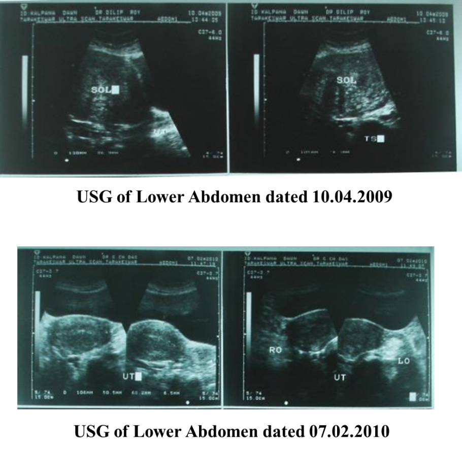
KD, 43 yrs old married women came to our clinic on 4th of May 2009 for the treatment of heavy menorrhagia with lower abdominal pain.
The USG of lower abdomen dated 10th of April 2009 showed “… a large SOL (10.5 cm x 7.4 cm) from fundus of uterus…”
After taking our specific medicines her lower abdominal pain is totally gone and menorrhagia is also controlled within 3 months.
The follow up USG of lower abdomen dated 7th of February 2010 showed “… as compared to the previous USG done on 10th of April 2009 the fundal SOL is not longer seen at present …”
Gall Bladder Stone Case
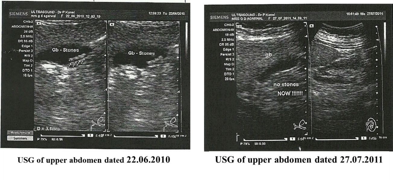
GDA, 62 years aged women came to our clinic on 1st of July 2010 with complaints of pain in right upper abdomen, gas in abdomen and heaviness in abdomen.
Ultrasonography of upper abdomen done on 22nd of June 2010 showed “…4-5 small calculi amid the lumen . Size of calculi = 08 - 09 mm…”
After taking our medicines her symptoms have improved within 3-4 months.
Follow up Ultrasonography of Upper abdomen done on dated 27th of July 2011 showed “…with comparison with the previous USG dated 22nd of June 2010, there is no evidence of cholelithiasis amid of the gallbladder. The gallbladder appears normal…”.
Gall Bladder Stone Case
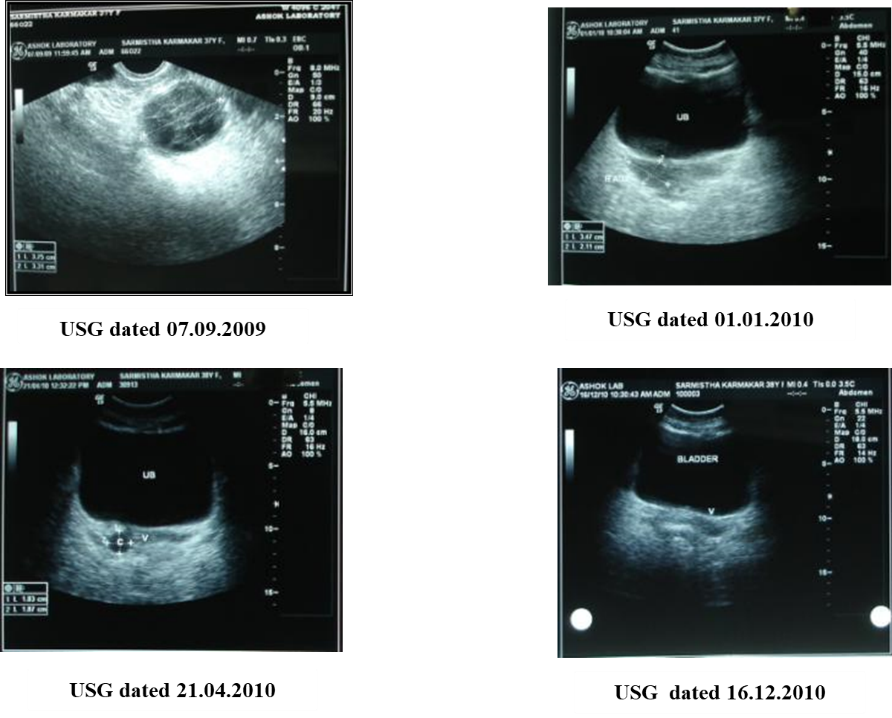
SK 37-year-old married lady came to us on 16th of September 2009 for the treatment of pain in her lower abdomen.
Before she visited us, the initial observation is
The U.S.G report of her lower abdomen dated 7th of September 2009 stated: Right adnexal complex S.O.L (8.6 cm X 4.4 cm) -? Loculated collection possibility of right ovarian S.O.L can not be ruled out. Mild free fluid in POD and also right iliac fossa region.
After taking our medicine, her abdomen pain was decreased.
Follow up U.S.G of lower abdomen report of 1st of January 2010 showed right ovarian cyst (3.5 cm x 2cm). Marked regression in size of the cystic lesion seen on comparing with study of 7th of September 2009.
She feels much better symptomatically and continues with the same medicines as prescribed.
Another follow up U.S.G. of lower abdomen report of 21st April 2010 showed further regression of the cystic lesion. The cyst now measures (1.8 cm x 1.8 cm.)
Another follow up U.S.G. report of her lower abdomen of 16th of December 2010 showed The cystic lesion seen in previous USG dated 21st of April 2010 has resolved completely.