Esophageal Carcinoma Case study 2
Mr. S D, male, aged 75 years was suffering for 2 months with difficulty in swallowing food, heartburn and belching, when he came to us for his treatment on 16th of December 1996.
Clinically the patient presented with dysphagia, heartburn and belching.

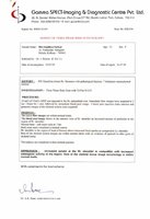
Endoscopy done on 29th of November 1996 showed "…GE junction at 40cm. At 18 cm. is a growth extending upto 22cm. causing luminal narrowing".
Biopsy dated 6th of December 1996, "…section shows moderately differentiated Squamous Cell Carcinoma".
After undergoing treatment from us with the medicine Condurango 30c 2 drops twice daily, the patients symptoms are gone within 2 months.
Now the patient is feeling much better. He is keeping good health and does not complain of dysphagia any more
Post treatment barium swallow X-ray of oesophagus dated 12th of July 1997 showed-"… there is considerable improvement in the patency of the oesophagus".
Esophageal Carcinoma Case study 2
M.R.I. of shoulder done on 06.07.2005 “ A large expansile osteolytic mass is seen in metaphysis and part of greater tuberosity of right humerus. There is extension to the paraosteal soft tissues.
Bone scan done on 26.07.2005 – Increase uptake at right shoulder is compatible with increase osteogenic activity in the region. Rest of the skeletal tissue image morphology is within normal limits.
C.T. guided F.N.A. of the lytic lesion in the humerus done on 28.06.2005 – The overall cytomorphological features are of malignant tumour of messenchymal origin.
The possibility of soft tissue sarcoma like synovial sarcoma with secondary involvement of the bone has been favoured.
Review report of the above slide on 04.07.2005 by Chittaranjan National Cancer Institute – “Impression :- The features are of malignant mesenchymal tumour – Pleomorphic sarcoma possibility of primary bone tumour is to be considered.”
Observations during treatment:
X-Ray right shoulder done on 18.02.2006 – Much improvement is seen of the osteolytic lesion in the upper part of the right humerus.
X-Ray right shoulder done on 01.02.2007- Bone destruction at upper 3rd of right humerus with soft tissue swelling.
Complication during treatment if any: Nil
Summary:
33 years aged young gentle lady came to us from remote village of Bangladesh on 22.07.2005 presented with Pain in and swelling in the right side of the shoulder since last 1½ months. As per her initial observations, M.R.I. of shoulder done on 06.07.2005 “ A large expansile osteolytic mass is seen in metaphysis and part of greater tuberosity of right humerus. There is extension to the paraosteal soft tissues.
Bone scan dose on 26.07.2005 – Increase uptake at right shoulder is compatible with increase osteogenic activity in the region. Rest of the skeletal tissue image morphology is within normal limits.
C.T. guided F.N.A. of the lytic lesion in the humerus done on 28.06.2005 – The overall cytomorphological features are of malignant tumour of messenchymal origin.
The possibility of soft tissue sarcoma like synovial sarcoma with secondary involvement of the bone has been favoured.
Review report of the above slide on 04.07.2005 by Chittaranjan National Cancer Institute – “Impression :- The features are of malignant mesenchymal tumour – Pleomorphic sarcoma possibility of primary bone tumour is to be considered.” Clinically patient feels much better and no swelling and pain in right shoulder area after 8 – 9 months of our medication. During her treatment X-Ray right shoulder done on 18.02.2006 – Much improvement is seen of the osteolytic lesion in the upper part of the right humerus. Again X-Ray right shoulder done on 01.02.2007- Bone destruction at upper 3rd of right humerus with soft tissue swelling. Patient feels much better and continuing medicines for further improvement.